What do these thick iron mats look like under the microscope? Here are three characteristic structures made by various iron-oxidizing bacteria that we’ve collected at Lo’ihi in the past few days.
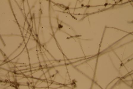
This sample was pulled from the fluffy “veil” that sometimes coats the wall of iron oxides. The tubular sheaths are formed by the bacteria cells as they grow. In fresh live cultures, bacterial cells can be seen lined up in a row within the walls of the sheaths.
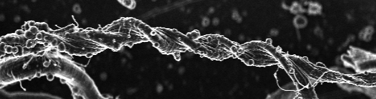
 This next sample was taken from a deeper, and most likely older section of the mat. It contains twisted stalks. In the case of stalks, we find individual cells at one end, with the stalk streaming behind like a tail.
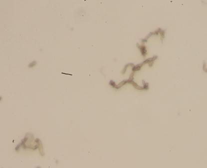
 We call the bacteria in the next image”Y-Guys” because of the branching patterns they make. You might find us doing the y-guy dance when we come across these guys under the microscope.
Check back soon to hear more about the samples we’re collecting!
-Anna Leavitt, Bigelow
Photos: Anna Leavitt

nice images Anna, will we get to see a video of the scientists doing the Y-guy dance….
Unfortunately we can’t get video up on the blog, but I did find this…http://earthref.org/galleries/2008/FEMO/day12/IMG_5683.html
🙂
ha ha ha, I had forgotten about that. I that that is the first move in the sequences too.
Pingback: Day 12: The Emerson Lab–cassettes, cultures, and interconnections | zetahunters